


비후성 심근병(hypertrophic cardiomyopathy) 사례, 심전도 이상에서 추가 평가
- Byoung-Yeon Jun

- 2020년 7월 17일
- 2분 분량
상기 환자 심초음파, 폐기능 검사, 트레드밀 (심장부하검사 시행함)
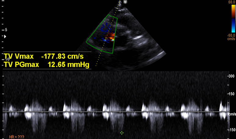
폐동맥 고혈압은 없으며

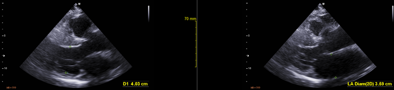
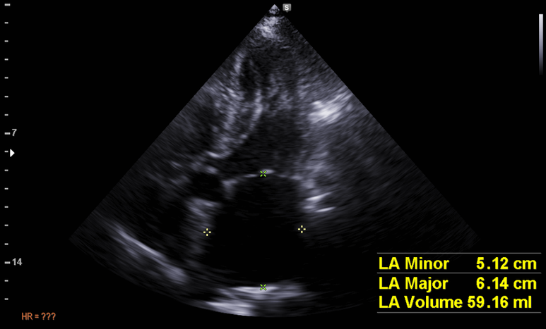
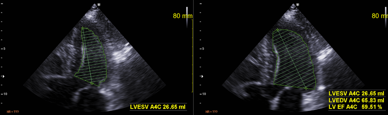
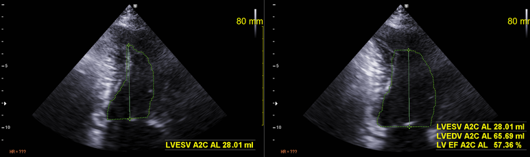
좌심실 부피는 40.8 ml/m2 으로 중등도의 확장

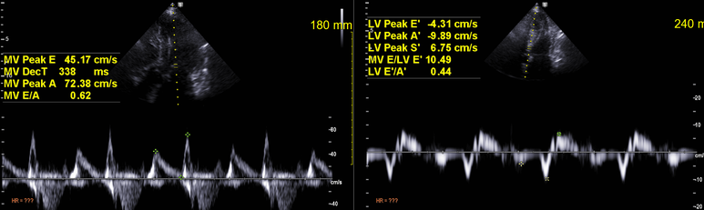
restrictive pattern의 이완기 장애
수축기 기능은 정상범위
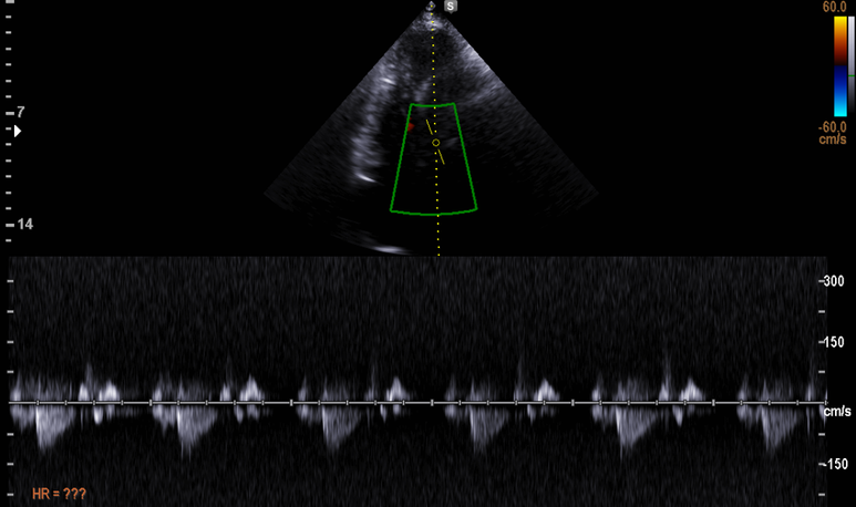
대동맥 출구에 증가된 혈류속도는 없으며

특히 좌심실 전벽에 2.3cm에 달하는 비후소견이 관찰됨
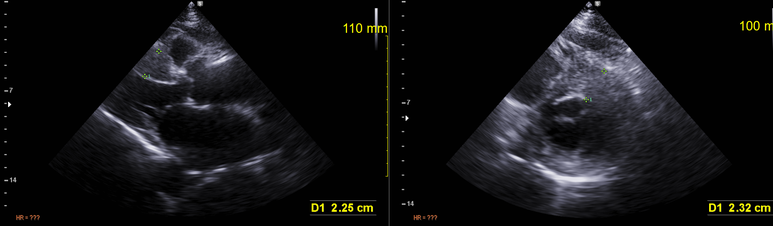
심실중격은 1.6cm정도, reverse curvature의 중격 모양을 보여주고 있다.

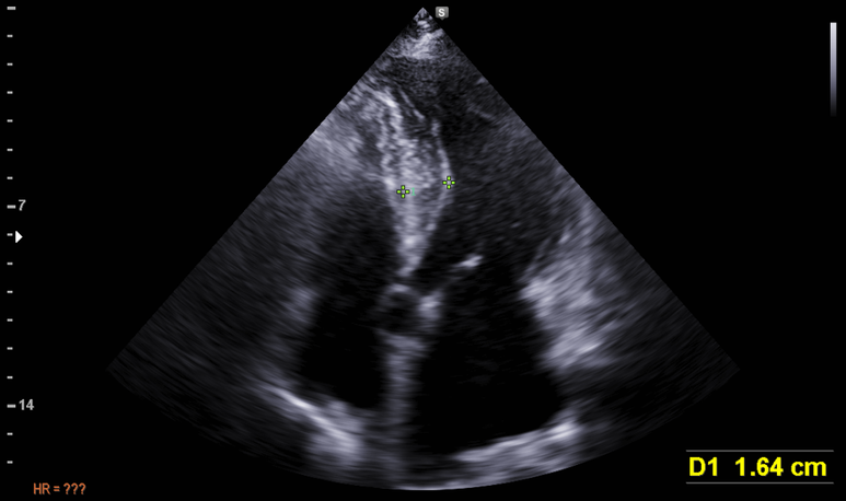


좌심실 비대, concentric hypertrophy (157g/m2)
이전 심전도를 다시 보면 이른 RS 전환 이외에 다음 아래의 소견을 볼 수 있다. 두번째 예시에서는 이른 RS전환 소견도 관찰할 수 있다.
<비후성 심근병 심전도 예시>

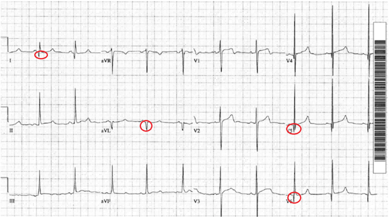

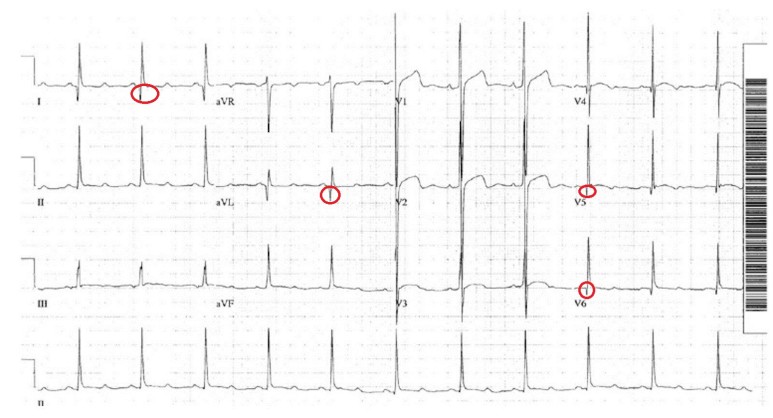
Classic HCM pattern with evidence of septal hypertrophy 심실중격 비대를 시사하는 전형적인 심전도 소견
1. Voltage criteria for left ventricular hypertrophy.
2. Deep narrow Q waves < 40 ms wide in the lateral leads I, aVL and V5-6.


Various anatomic forms of hypertrophic cardiomyopathy and their relation to genotype status. Studies have demonstrated a higher rate of genopositivity in the setting of specific morphologic subtypes. The yield from testing was highest in those patients with either a reverse or neutral septal morphology, in comparison with a low yield in those patients with a sigmoid septum or apical variant. , * and ** represent numbers derived from two major studies
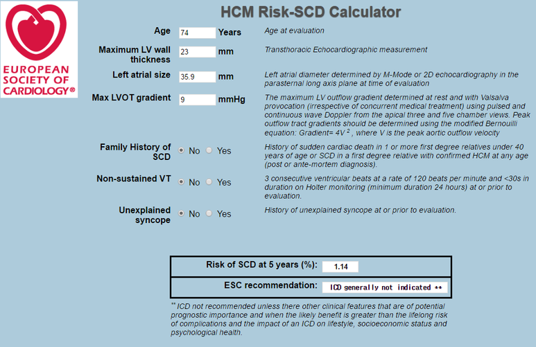
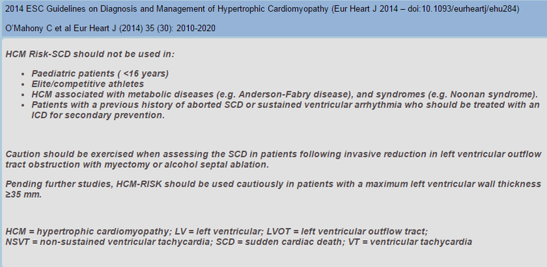
혈압 및 심혈관 위험인자 조절 유지하고 심초음파 추적 계획 - 중격 비대 변화, 좌심실 출구 협착 발생 여부 추적
증상 발생시 betablocker(안정시 심박수 50/min), nitrate/diuretics 추가할지 결정
부정맥 여부 확인 위해 holter계획, 1촌 가족의 심초음파 계획


Reproduced with permission. Circulation. 2011;124 iss 24: e783-831. ©2015 American Heart Association, Inc.


동대문구 답십리 우리안애, 우리안愛 내과, 건강검진 클리닉 내과 전문의 전병연


Comments