초진환자에서 간경화 설명의 어려움, 복수도 있으니 비대상성에 해당해도... - 동대문구 답십리, 용두동, 우리안애 우리안愛 내과
- Byoung-Yeon Jun

- 2023년 10월 16일
- 4분 분량
40대 초반 여자, 초진
알콜병원 입원 하여 금주중
; 입원시 타병원/의원 진료시에는 의뢰서를 첨부해서 방문해야하며 본인부담 100%로 처리 후 입원 병원에서 청구하여 정산해야한다.
의뢰서 줄 때 입원병원이면 안내를 잘 해줘야 하는데 모른척을 하고 보내는지...
계획한 초음파 비용이 많아서 다른날 다시 방문하였다.
정강이에 부종 때문에 평가하러 왔는데 알콜 질환을 배경으로 하니 간경화 여부, 복수 여부가 관찰의 주안점이 된다.
약간의 함요부종은 확인되는데 심하지는 않다.
과거 경기도에서 초음파시 간이 부었다고 들었다? ... 이해를 위한 일반적인 설명이겠으나... 부정확하다.
비장이 크다고도 들었다.
약 4~5년 전부터 간헐적 폭음.... 640 ml 소주 3병을 1주일간 마시기도 하고...
초음파 시행
좌측간의 외측구획과 1번 구획 사이가 벌어지고 좌측간 뒤쪽의 결절성 변화가 뚜렷

B형간염 초진 환자들에서 이런 모습이면 설명하기가 어렵다 (혹은 이해하기 어려워한다.)
간문맥의 직경이 확장되어 있다. > 12 mm

비슷한 직경의 간문맥, 담낭벽 비후/복수의 정도, 다리 함요부종이 더 심하고 뚜렷했던 경우
담낭벽도 경미하게 전반적으로 두꺼운데... 혈액검사에서 알부민이 낮지 않을까 추정한다.
현재 입원병원 주치의가 알고 있을 것이며...

복수가 더 심한 경우 담낭벽 비후가 더 뚜렷하다.
아래 경우는 알콜성 간경화로서 다리의 부종이 윗 환자보다 훨씬 뚜렷하다. 아래 나오겠지만 복수의 차이도 있는데 모든 소견에서 차이가 연관된다.
전반적으로 간의 실질이 거친것이 뚜렷, 진행된 섬유화를 시사한다.

간실질이 거친 배경에서 (10년간 금주했음에도) 간암의 발견
B형 간염환자, 간경화로 판단
우엽 후반부에 결절 변화는 매우 크게 관찰된다. Right posterior hepatic notch sign

금주하고 있으나 초음파 소견은 간경화가 뚜렷한 모습
그러나, 간우엽의 전반부에 요철변화는 뚜렷하지 않다.

좌엽의 4번구획 (medial segment) 와 2/3번구회 (lateral segment) 사이에 요철변화

비장정맥의 측부 순환은 없다.


알콜성 간경화에서 측부순환
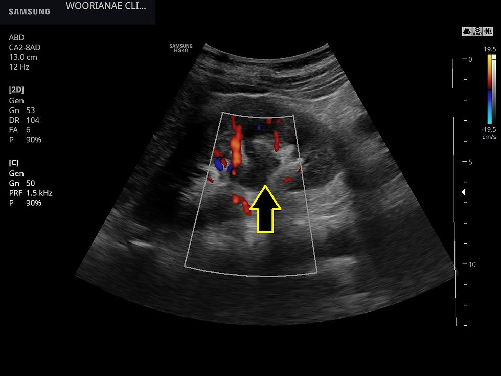
양측에 수신증이 뚜렷하게 관찰되었다. 원인은?


The cause of urinary obstruction can be broadly classified as intrinsic and extrinsic compression. Causes of intrinsic obstruction include renal stones, malignancy, ureteropelvic junction stenosis, ureteral strictures from prior inflammation, renal cysts, posterior urethral valves, benign prostatic hyperplasia, and neurogenic bladder, etc. Causes of extrinsic compression include pregnancy, peripelvic cysts, retrocaval ureter, malignancy, trauma, retroperitoneal fibrosis, and prostate abscess, etc. https://www.ncbi.nlm.nih.gov/books/NBK563217/ 임상적으로 일측, 혈뇨를 동반한 옆구리 통증의 상황 (요로결석) 은 아니며... https://blog.naver.com/ejercicio/221693740462
소량의 복수가 방광 좌우, 위쪽으로 관찰된다.


독성 간염, 소량의 복수, 간주변 및 방광주변
알콜성 간염, 다량의 복수 https://blog.naver.com/ejercicio/222115528141
비대상성 간경화, 복수 https://blog.naver.com/ejercicio/222549789460
우측간의 뒷면을 다시 관찰함
결절 변화가 뚜렷하다.


종합하면 간경화 모습인데
혈액결과는 어떠했을까? 혈소판 감소, 알부민 감소, PT 의 연장?
다른 임상 정보를 모른체 초음파 만으로 간경화 설명하기는 듣는 사람이 이다/아니다의 이분법으로 알아듣기 때문에 조심스러울 수도 있다. 사실은 이행하는 경과임에도... (섬유화... 심한섬유화...간경화...)
복수를 제외하더라도 종합하면 충분히 결론 지을 수가 있고 복수를 고려하면 비대상적 변화이기 때문에 더욱 뚜렷하다고 할 수 있다.
1. 복수의 다른 원인을 배제하기 위해 CT의 필요성? 간에 결절은 없지만, CT가 초음파보다 우선한다고 할 수 없지만 간경화 평가를 위해 시행?
2. 수신증 평가를 위해 CT는 추가적으로 필요
3. 알콜병원에서 금주한지 수주가 되어 가니.. 퇴원후 재범 (recidivism) 이 아니길 기대하며, 과거 3차병원 평가력이 있어서 방문하기로 하였는데 술을 계속 마신다면 병의 진행에서 어느 병원을 가도 마찬가지.. 따라서 강제성이 있는 알콜병원에서의 계획 및 3차 병원 평가 및 관리 (복수/부종 조절을 위한 이뇨제의 처방등) 의 균형을 잘 잡으면 좋겠다; 경도의 부종은 복수가 생긴 비대상성 간경화로 설명하면 된다.
Diagnosis of liver fibrosis and cirrhosis
Histology, biology, and transient elastography Histology remains the reference standard for the diagnosis of fibrosis and cirrhosis. However, there are several concerns related to this technique. First, it is an invasive technique with potential complications and therefore not conducive to follow-up. Second, the size of the sample is very small, and is therefore possibly not representative of the entire liver lesion. Finally, the reproducibility of the Metavir score is low. Biopsy is important for assessing associated lesions, such as inflammation or balloonization of cells, but its use in fibrosis quantification is limited. At present, in clinical practice, the diagnosis of liver fibrosis and cirrhosis is mainly based on clinical, biological, and elastographic data [6,51].
Although the radiologic features of liver fibrosis and cirrhosis can be found on CT and MRI scans, ultrasound is the main tool used in clinical practice for the diagnosis of fibrosis and cirrhosis [52]. Among the previously listed signs, the nodular aspect of the liver surfaces, increased spleen length, and demodulation of the hepatic vein appear to be most accurate in the diagnosis of severe fibrosis. However, all the other listed signs could be encountered in severe fibrosis or cirrhosis and could contribute to the diagnosis. It is important to keep in mind that there is no specific sign that makes it possible to diagnose severe fibrosis, since diagnosis is usually based on an association of signs [12]. 하나의 소견이 특이적으로 심한 섬유화를 진단할 수는 없다. 진단은 연관된 여러 사인/소견 에 종합하여 결정해야 한다. In literature, the accuracy of sonographic and Doppler measurements for the detection by experienced physicians of cirrhosis are estimated to be between 82% and 88% [5,11,53]. Among the different elastography techniques recently proposed by manufacturers of ultrasound imaging devices, two are currently well evaluated in literature: ARFI (acoustic radiation force impulse) by Siemens and SWE (shear-wave elastography) by Supersonic Imagine. The sensitivity of these techniques for the diagnosis of severe fibrosis and cirrhosis varies from 79% to 97% and 81% to 95%, respectively, and the specificity varies from 81% to 87% and 77% to 96%, respectively, according to various studies [35,37]. The cutoff values of stiffness used for severe fibrosis are 1.5 m/s for ARFI and 8.7—8.9 kPa for SWE. For cirrhosis, the values are 1.61 m/s for ARFI and 10.4—10.7 for SWE [35,37]. Differences in cutoffs are explained by differences between populations studied and between techniques [44].
<elastography> - 간섬유화, 간탄성도 검사도 수치로 평가 정보를 주지만 다른 소견이 간경화/진행한 섬유화인데 그 수치가 다른 사인들을 기각시키면 안되겠다. 참조하여 민감도와 특이도를 향상시켜준다고 한다.
고가의 장비라서 의원급에서는 시행하는 곳이 거의 없다.
동대문구 답십리 우리안애, 우리안愛 내과, 건강검진 클리닉 내과 전문의 전병연
#동대문구내과 #성동구내과 #광진구내과 #답십리내과 #장안동내과 #용답동내과 #청량리내과 #추천 #왕십리역 #장한평역 #답십리사거리 #촬영소사거리 #전농동내과 #내과 #건강검진 #위내시경 #대장내시경 #갑상선초음파 #복부초음파 #경동맥초음파 #심장초음파 #암검진 #래미안위브아파트 #엘림스퀘어 #두산아파트 #동아아파트 #한양아파트 #동답한신아파트 #두산위브아파트 #힐스테이트청계아파트 #래미안미드카운티 #청솔우성 #래미안미드카운티


Comments